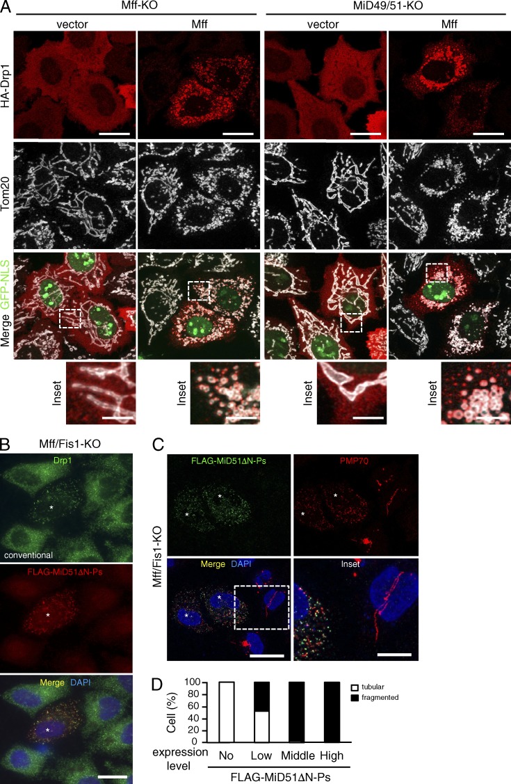
Figure 3.
Mff and MiD51 independently mediate membrane fission. (A) Confocal images of the indicated cells cotransfected with HA-Drp1 and empty vector or pMff-IRES-GFP-NLS. Cells were immunostained with the indicated antibodies. Bottom, magnified images of the boxed regions. Bars: 10 µm; (magnification) 2.5 µm. (B–D) Mff/Fis1-KO cells were transfected with FLAG-MiD51ΔN-Ps and subjected to immunostaining with the indicated antibodies. Conventional (B) and confocal images (C) are shown. Asterisks, FLAG-MiD51ΔN-Ps expressing cells. Magnified image of the boxed region is shown as inset. Bars: 20 µm; (magnification) 10 µm. Nuclei were stained with DAPI in B and C. (D) Percentages of cells with indicated peroxisomal morphologies in cells (n = 100).

