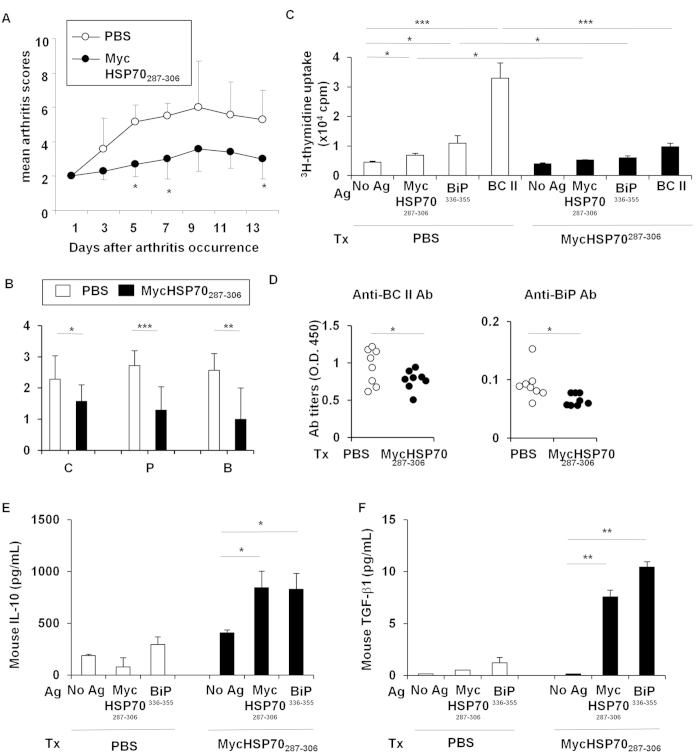
Figure 6. Oral tolerance of MycHSP70287–306 peptides in a collagen-induced arthritis model.
(A,B) The MycHSP70287–306 peptide or control (PBS alone) was orally administered to bovine type II collagen-immunized DBA/1 J mice after the occurrence of CIA for 5 continuous days (n = 12). Mice were sacrificed on day 35. Arthritis scores were measured by finger and paw swelling, and histological scores were measured by cell infiltration (C), pannus formation (P), and bone destruction (B). (C) Splenic CD4+ T cell proliferation from CIA-induced DBA/1 J mice in response to a recall antigen (Ag) stimulation (MycHSP70287–306 and BiP336–355 epitopes, and heat-denatured bovine type II collagen (BC II)). Proliferation was determined by3-H thymidine incorporation. (D) Serum anti-BiP and anti-BC II antibody titers were measured by ELISA. (E,F) Mouse IL-10 and TGF-beta1 concentrations in the splenic CD4+ T cell culture were measured by ELISA. Data were representative of at least three independent trials. *p < 0.05, **p < 0.01, ***p < 0.001.

