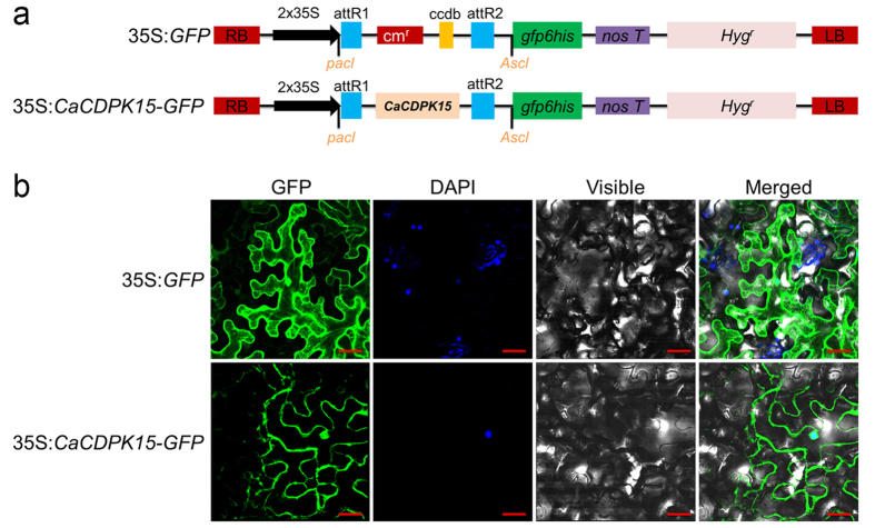
Figure 2. Subcellular location of CaCDPK15.
(a) Schematic representations of the vector constructs of 35S:GFP and 35S:CaCDPK15-GFP. (b) GFP fusion protein of CaCDPK15 was localized in the cytomembrane and nucleus of N. benthamiana cells. The plant nuclei were stained with DAPI. Images were taken by using Leica confocal microscopy at 48 hours post agroinfiltration (GFP fluorescence, green; DAPI fluorescence, blue; visible, visible light image; merged, merged images of above three images). Bars = 200 μm.

