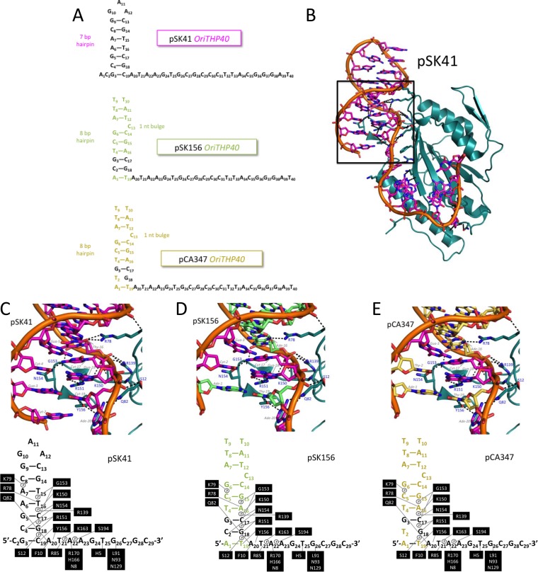
FIG 3.
Modeled structures of pSK41, pSK156, and pCA347 oriT regions. (A) Schematic of the pSK41, pSK156, and pCA347 oligonucleotides used in these studies. Colored nucleotides indicate differences in sequence from the pSK41 oriT. (B) Relaxase domain of NES in complex with pSK41 oriT. The box shows the region focused on for panels C to E. (C) Contacts between NES relaxase domain hairpin loop 1 and 2 amino acids and the pSK41 oriT nucleotide. (D) Contacts between NES relaxase domain hairpin loop 1 and 2 amino acids and the modeled pSK156 oriT nucleotide. Green nucleotides differ from the pSK41 oriT region. (E) Contacts between NES relaxase domain hairpin loop 1 and 2 amino acids and the modeled pCA347 oriT nucleotide. Gold nucleotides differ from the pSK41 oriT region.

