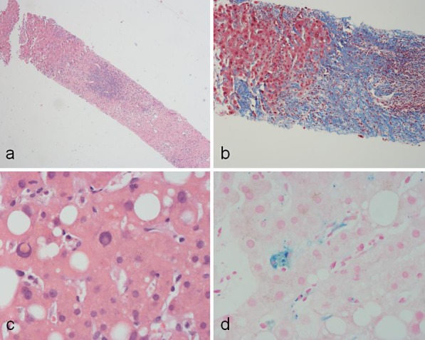
Fig. 2.

Needle core biopsy of the liver taken 47 months after 90Y administration. a There is a large band of chronically inflamed fibrous tissue, highly suggestive of cirrhosis. HE. ×10. b This is highlighted by Masson trichrome staining, demonstrating chronically inflamed fibrous tissue (blue), highly suggestive of cirrhosis. ×10. c The nonfibrotic liver showed macrovesicular steatosis with mildly active steatohepatitis. HE. ×40. d On the other hand, sinusoidal Kupffer cells showed a mild degree of hemosiderosis. Perls iron stain. ×40.
