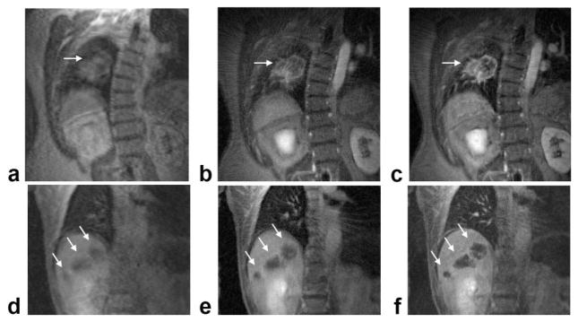
FIG. 6.
Comparison of image quality using the proposed golden-angle radial scheme with conventional Cartesian acquisition. a: Cartesian DCE-MRI image of the lung lesion. b,c: Radial images of the lesion, reconstructed from either 550 consecutive views (b) or from 10 consecutive 55-view motion-compensated end-expiratory segments (c). d–f: Corresponding images from the liver tumor patient. For this patient, the radial images were reconstructed from either 340 consecutive views (e) or 10 consecutive 34-view end-expiratory segments (f). Lesions are indicated by arrows.

