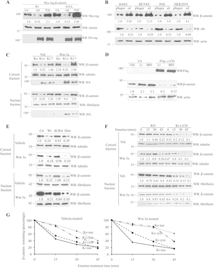
FIGURE 3.
c-Cbl destabilizes β-catenin during both the phases of Wnt signaling. A, c-Cbl down-regulates β-catenin. HUVECs stably expressing control vector (Ctr), HA-tagged c-Cbl (c), or c-Cbl-70Z (70Z) were transiently transfected with various constructs of Myc-tagged β-catenin. Digitonin-extracted cytosol fractions were immunoblotted using HA, Myc, and actin antibodies. Representative immunoblot from two experiments is shown. B, c-Cbl silencing in ECs up-regulates β-catenin. Lysates of cells stably silenced with control (pSup) or c-Cbl (sil) retrovirus were probed with β-catenin, c-Cbl, and actin antibodies. Representative immunoblot from three experiments is shown. C, increased β-catenin levels in c-Cbl knock out (KO) ECs. Lysates from ECs from c-Cbl KO or with knock-in (Ki) with HA-tagged c-Cbl (Ki-c) or c-Cbl-70Z (Ki-7) and pretreated with vehicle (veh) or Wnt3a 100 ng/ml were fractionated and immunoblotted using β-catenin, HA, and actin antibodies. Representative immunoblot from three experiments is shown. D, c-Cbl regulates β-catenin stabilized by inhibition of GSK-3β. ECs stably expressing control (Ctr) or FLAG-c-Cbl were serum-starved and treated with vehicle (Veh) or lithium (10 mm) and BIO (100 nm) for 12 h. Whole cell lysates were probed for β-catenin, FLAG, and actin antibodies. β-Catenin bands were normalized to actin using ImageJ. Representative immunoblot from two experiments is shown. E, c-Cbl dimerization-dependent regulation of β-catenin. ECs stably expressing FLAG-tagged c-Cbl constructs were serum-starved and stimulated with Wnt3a (50 ng/ml) followed by subcellular fractionation. The lysates were probed for β-catenin. Tubulin and fibrillarin served as markers of cytosolic and nuclear fractions and as loading controls, respectively. Representative immunoblot from two experiments is shown. F, c-Cbl destabilizes endogenous β-catenin in both the phases of Wnt signaling. Lysates from c-Cbl KO or KI ECs serum-starved for 16 h and pretreated with Wnt3a (100 ng/ml) for 3 h and 20 μm emetine were fractionated at different time intervals and immunoblotted for endogenous β-catenin. Tubulin and fibrillarin served as cytosolic and nuclear markers, respectively, and loading controls. Densitometric values are normalized loading controls. Representative immunoblot of three experiments is shown. G, percent β-catenin remaining was analyzed by densitometry after normalizing to tubulin and fibrillarin, respectively. Graph shows mean result of three experiments. Error bars, S.E. WB, Western blot.

