
Figure 3.
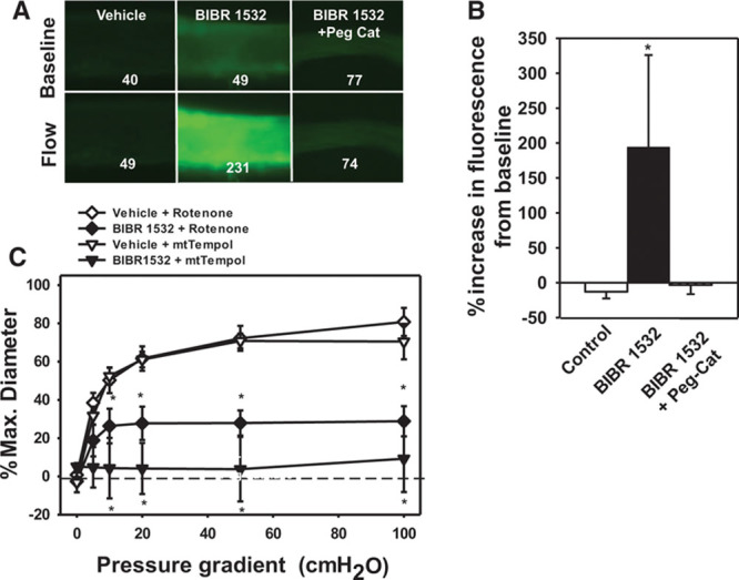
Mitochondrial hydrogen peroxide (H2O2)–mediated flow-mediated dilation (FMD) after inhibition of telomerase. H2O2 levels were analyzed using the H2O2-specific florescent probe PYI (peroxy yellow 1) targeted to mitochondria (MitoPYI) in vessels from non–coronary artery disease (CAD) subjects. A, Representative image; numbers represent florescent intensity above background. B, Summary of florescence intensity at 5 minutes after initiation of flow. Specificity of the probe for H2O2 was confirmed using polyethylene glycol-catalase (Peg-Catalase). C, An inhibitor of electron transport chain complex I (rotenone) or a mitochondrial-targeted reactive oxygen species (ROS) scavenger (MitoTempol) inhibited FMD after telomerase inhibition. N=4. *P<0.05 2-way analysis of variance (ANOVA) RM (dose response curve) or t test (fluorescence data) with Tukey post hoc.