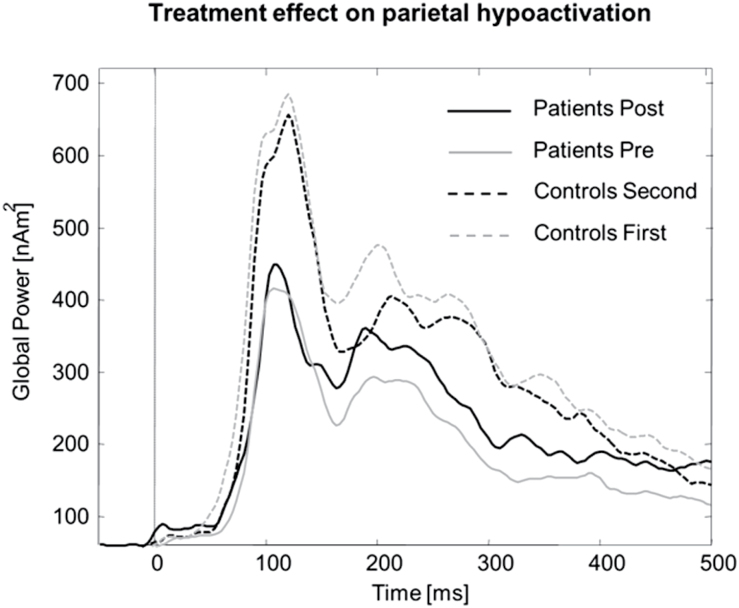
Figure 4.
Time course of parietal cortex activity for 15 major depressive disorder patients who participated in both the pre- and post-therapy magnetoencephalography (MEG) sessions, and their matched healthy controls in their first and second MEG sessions. While parietal activation, most probably due to habituation and reduced vigilance, decreased in the control group, patients showed increased parietal activity after therapy in direction normalization.

