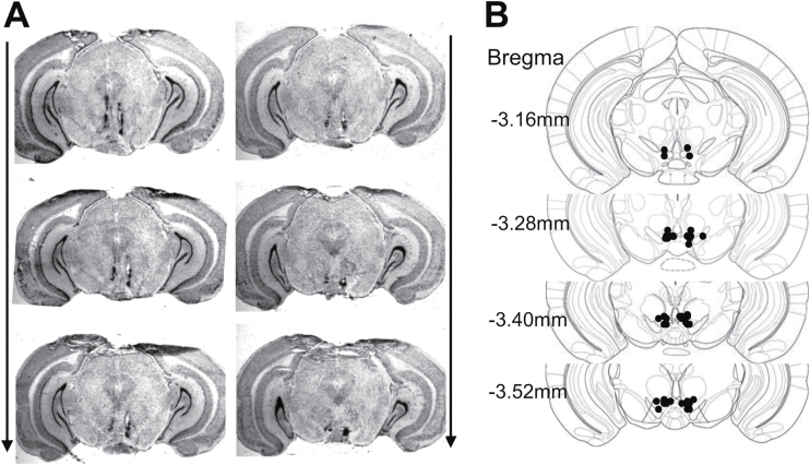
Figure 5.
Histological verification of intra-ventral tegmental area (VTA) injection sites. (A) Representative images showing the deposition sites within the VTA. Arrows indicated the rostral-caudal sequence of the coronal sections. (B) Schematic illustration of bilateral injection sites in the VTA. The drawings of coronal sections were derived from the atlas of Paxinos and Franklin (2001).

