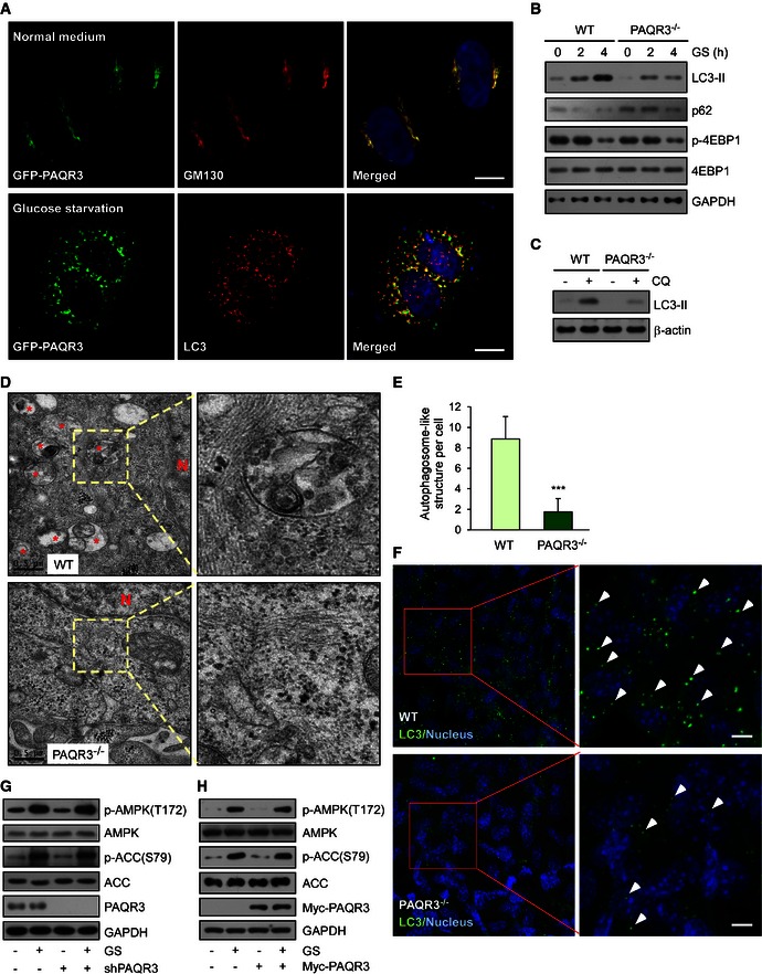
Figure 1. PAQR3 regulates autophagosome formation without altering the activities of AMPK and mTOR .

-
AGFP‐PAQR3‐transfected HeLa cells were fixed for immunofluorescence staining with the indicated antibodies before or after glucose starvation (GS) for 4 h. The nuclei were stained with Hoechst 33342. Scale bar: 10 μm.
-
BImmunoblotting (IB) analysis of wild‐type (WT) and PAQR3‐deficient MEFs after GS for different times as indicated.
-
CWT and PAQR3‐deficient MEFs were treated with 80 mM chloroquine (CQ) for 2 h to induce LC3‐II accumulation. Whole‐cell lysates were harvested for IB analysis of LC3‐II.
-
DAutophagosome‐like structures in WT and PAQR3‐deleted MEFs were detected after GS for 4 h by transmission electron microscopy. * indicates autophagosome‐like structures. N, nucleus.
-
EQuantitative analysis of autophagic vacuoles in transmission electron microscopy images. Thirty cells were quantified from each independent experiment, which was repeated for three times with similar results. Values are presented as mean ± SD, ***P < 0.001.
-
FImmunofluorescence staining of LC3 in WT or PAQR3‐deficient MEFs after GS for 4 h. The arrowheads indicate representative LC3‐positive puncta. Scale bar: 10 μm.
-
G, HHeLa cells were infected by PAQR3 knockdown or overexpression of lentivirus together with their respective control virus. After GS for 1 h, the cell lysates were harvested for IB.
