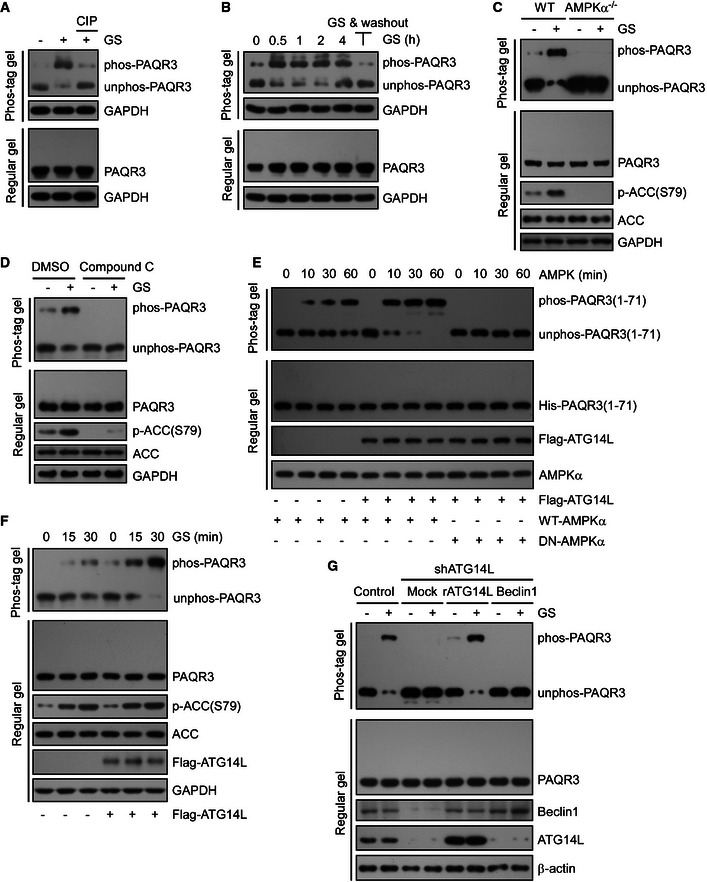
HeLa cells were cultured in normal medium (NM) or glucose starvation (GS) for 4 h. The starved lysates were treated with calf intestinal phosphatase (CIP) to remove phosphate groups and analyzed by immunoblotting (IB) with Phos‐tag gel or regular SDS–PAGE.
The cell lysates from HeLa cells were collected and analyzed after GS for the indicated time (lane 1–5), or GS for 2 h followed by 2‐h chase in NM (lane 6). The cell lysates were subjected to Phos‐tag gel and regular SDS–PAGE, respectively.
WT or AMPKα1/2 double knockout MEFs were incubated under NM or GS for 4 h, followed by IB with Phos‐tag gel or regular SDS–PAGE.
HeLa cells were incubated with compound C (20 mM, 30 min) with or without GS, followed by IB with Phos‐tag gel or regular SDS–PAGE.
Bacterial purified His‐tagged NH
2‐terminal 71 amino acid of PAQR3 was incubated with or without purified Flag‐tagged ATG14L. Then, the complexes were treated with WT AMPK or DN‐AMPK (dominant negative AMPK) for the indicated time in vitro. PAQR3 phosphorylation was examined by Phos‐tag gel.
HeLa cells infected with control or ATG14L‐expressing lentivirus were incubated under GS for different times as indicated. Whole‐cell lysates were analyzed by both Phos‐tag gel and regular SDS–PAGE.
HeLa cells were infected by lentivirus expressing control shRNA or ATG14L‐specific shRNA. Then, the ATG14L knockdown cells were stably re‐expressed with shRNA‐resistant ATG14L (rATG14L) or Beclin1 and incubated under NM or GS for 4 h. Whole‐cell lysates were analyzed by both Phos‐tag gel and regular SDS–PAGE.

