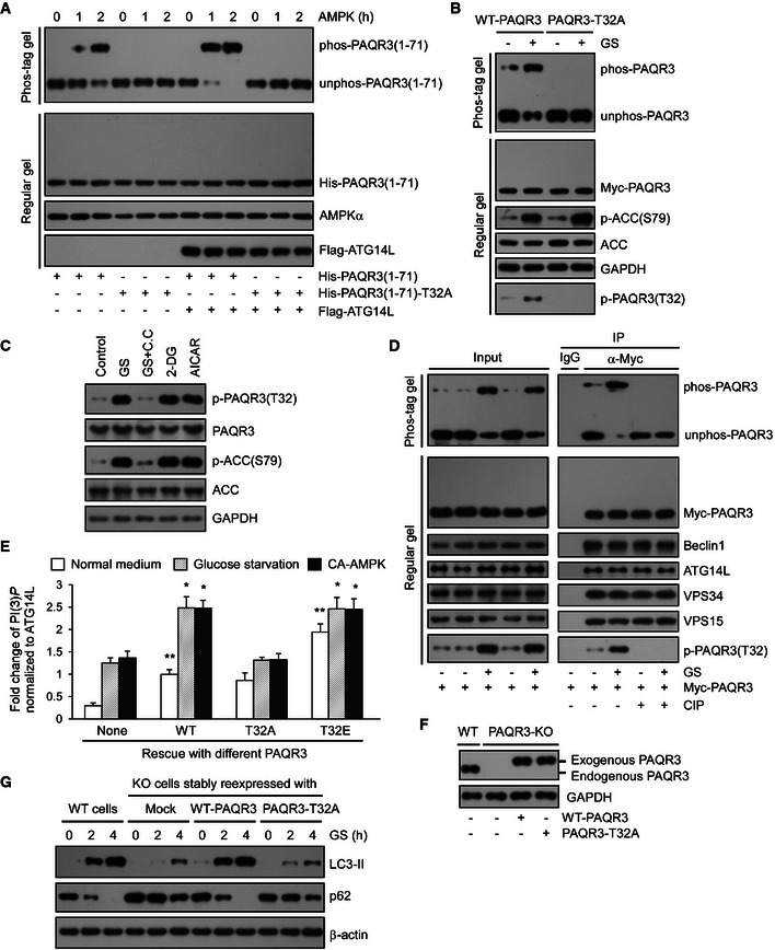
Figure 6. Phosphorylation of PAQR3 T32 is required for autophagy initiation upon glucose starvation.

- Bacterial purified His‐tagged WT or T32A NH 2‐terminal 71aa of PAQR3 was incubated with or without Flag‐tagged ATG14L purified from HEK293T cells. Then, the complexes were incubated with AMPK for the indicated time and subjected to Phos‐tag gel or regular SDS–PAGE.
- PAQR3‐deficient HeLa cells were transfected with WT or T32A mutants of PAQR3 plasmids. Twenty‐four hours later, the cells were incubated with normal medium (NM) or glucose starvation (GS) for 4 h, followed by immunoblotting (IB) in Phos‐tag gel or regular SDS–PAGE.
- HeLa cells were incubated with NM or GS for 1 h or treated with compound C (C.C, 20 mM, 30 min) prior to GS for 1 h. In parallel, 25 mM 2‐DG or 1 mM AICAR was added in NM for 1 h. Cell lysates were analyzed by IB.
- HEK293T cells were transfected with Myc‐tagged PAQR3. At 24 h after transfection, the cells were treated with GS for 4 h. Then, the cell lysates were used in IB and immunoprecipitation (IP) with the antibodies as indicated. The immunoprecipitates were treated with CIP to remove phosphate groups and analyzed by IB in Phos‐tag gel or regular SDS–PAGE.
- WT, phosphorylation defective (T32A), or phospho‐mimetic (T32E) PAQR3 were transfected in the presence or absence of constitutively active AMPK (CA‐AMPK) into PAQR3‐deficient HeLa cells as indicated. Twenty‐four hours later, the cells without AMPK transfection were treated with GS for 4 h. About 10% of the cell lysates were subjected to IB to detect PAQR3 expression level (shown in Appendix Fig S4A), and the remaining cell lysates were used to immunoprecipitate VPS34 complexes by ATG14L antibody, followed by VPS34 activity measurement by a quantitative PI(3)P ELISA. The PI(3)P level was normalized to the amount of ATG14L. Values are presented as mean ± SD (n = 5; *P < 0.05; **P < 0.01 as compared to the first group).
- PAQR3‐deficient HeLa cells were infected with lentivirus expressing WT or T32A PAQR3. Then, PAQR3 expression levels were examined by IB.
- Both WT HeLa cells and PAQR3 knockout HeLa cells infected with lentivirus expressing WT or T32A PAQR3 were treated with GS for 2 or 4 h, respectively. Cell lysates were analyzed by IB.
