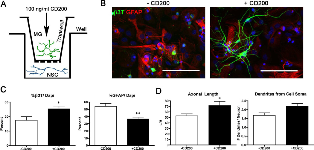
Figure 6. CD200-stimulated microglia increase neural differentiation of neural stem cells and Aβ phagocytosis in vitro.
(A) Scheme shows the experimental design for the in vitro co-culture study. Neural stem cells were co-cultured with CD200-pre-treated microglia in transwells for seven days. (B) Images of neural stem cells after co-culture with CD200-treated microglia stained with astrocytic marker GFAP (red) and mature neuronal marker β3T (green). Scale bars represent 100 µm. (C) Quantification of cellular differentiation was performed by counting the percentage of β3T+ or GFAP+ cells over total number of cells. (D) Neuronal complexity was determined by measuring axon length using NeuronJ and total number of dendrites emerging from the cell soma (n= ~50 per group). * and ** denote p<0.05 and 0.01, respectively, as determined by Student’s t-test. (E) Microglia were pre-treated with CD200 followed by incubation with oligomeric Aβ42 for 72 hours. Microglia were stained for Aβ (NU-1, green) and Hoechst 33342 (blue). Scale bar represents 100 µm. * and ** denotes p<0.05 and 0.01, respectively, as determined by Student’s t-test.

