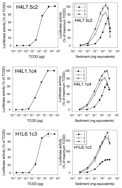
Figure 3.
Sediment extracts induce luciferase in a dilution-dependent manner in rat hepatoma G3 CALUX cells and previous generation mouse and rat hepatoma CALUX cells. Rat H4L7.5c2 G3 CALUX cells, rat H4L1.1c4 CALUX cells, or mouse H1L6.1c3 CALUX cells were incubated with TCDD or three different sediment extracts, and luciferase activity in cell lysates was measured as described under Materials and Methods. The sediment extracts studies correspond to those in positions 5A, 5C and 6E of the plates presented in Figure 2. Luciferase activity was expressed as a percent of maximum TCDD induction and values represent the mean ± SD of triplicate determinations after subtraction of the luciferase activity obtained in cells exposed to DMSO (for TCDD) or method blanks (for sediments). These data are representative of results from three separate experiments.

