Fig. 2.
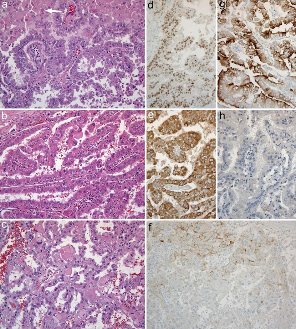
Tumor histology and immunohistochemistry a. Carcinoma from the left adrenal is composed of cells with eosinophilic cytoplasm, large nuclei, and prominent nuclei. Papillary architecture is shown at bottom, while cells at the top of the microscopic field have more abundant cytoplasm and some intracytoplasmic hyaline inclusions (arrow) b. Carcinoma from the liver metastasis has prominent papillary architecture and similar cytologic features; admixed neutrophils are present c. Carcinoma from the right adrenal is again similar, but there are scattered very large cytoplasmic hyaline inclusions in this tumor focus d. PAX8 immunohistochemical staining positive in tumor cells e. Racemase (AMACR, P504S) strongly diffusely positive in tumor cells f. Immunostaining for the von Hippel–Lindau protein essentially negative; there was only weak cell membrane, or apical staining focally within the carcinoma, as shown in the top of the panel. The expected normal staining pattern diffuses cytoplasmic and membrane staining (not shown). g. RCC monoclonal antibody staining positive with the expected apical membrane accentuation. h. SF-1 immunohistochemistry negative. Original magnifications: 200×

