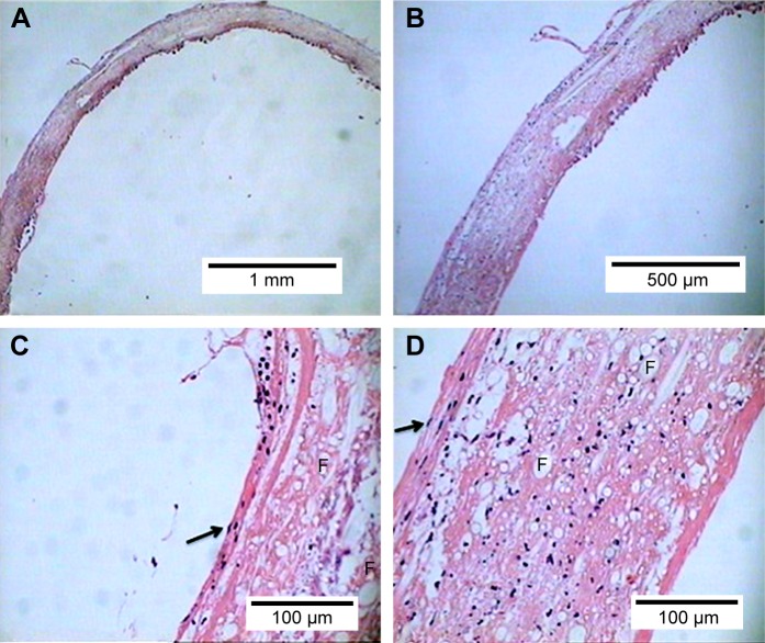
Figure 3.
Histological analysis. Hematoxylin–eosin staining. The scaffold appeared to be densely colonized by different cellular elements that progressively acquired different phenotypic characteristics according to the region of the TEVG in which they engrafted. (A) 5× magnification. (B) 10× magnification. (C) 40× magnification of the inner side of the TEVG. Note flat elongated cells with nucleus protruding in the lumen (arrow) organized in an endothelial-like fashion. (D) 40× magnification of the outer side of the TEVG. Note spindle-shaped cells reliably representing fibroblasts (arrow). F indicates fibers of polymer in both cross and long axis section.

