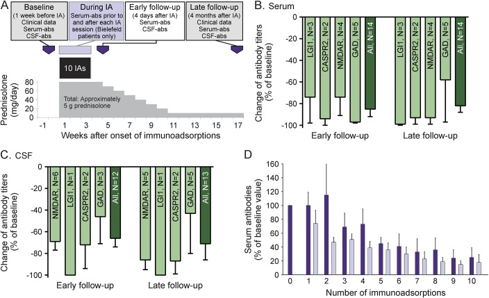
Figure 1. Standard treatment scheme and antibody titer changes.
(A) Schematic depiction of the standard treatment schedule. (B–D) Changes of antibody titers in serum (B) and CSF (C) at early and late follow-up in relation to the antibody targets (means with standard errors of the means). (D) Antibody titers of all patients (pooled) from Bielefeld-Bethel (n = 15, means and standard errors of the means) on the immunoadsorption days directly prior to (dark purple bars) and after the immunoadsorptions (light purple bars). This shows the known sawtooth configuration that reflects the antibody redistribution between the vascular and extravascular compartments. Even after the fifth session, there is ongoing reduction of antibodies. CASPR2 = contactin-associated protein-2; GAD = glutamic acid decarboxylase; IA = immunoadsorption; LGI1 = leucine-rich glioma inactivated protein 1; NMDAR = NMDA receptor.

