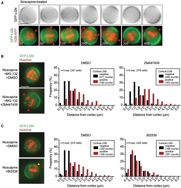
Figure 2. Plk1 restricts cortical LGN localization near misaligned chromosomes.

-
AHeLa cell stably expressing GFP‐LGN and transiently expressing RFP‐H2B was treated with noscapine to induce chromosome misalignments. Cells were imaged every 5 min. The time is relative to NEB. The red arrows indicate cortical regions where the chromosomes are in close proximity to the cell boundaries, accompanied by the displacement of LGN from the cortex. Scale bar represents 10 μm.
-
B, CSimilar assay as described in Fig EV3E was carried out in the presence of a small molecule inhibitor against Aurora B (ZM447439) in combination with the proteasome inhibitor MG‐132 to keep cells arrested in mitosis (B) and with an inhibitor against Plk1 (BI2536) (C). Drugs were added at the same timing as Hoechst, 30 min prior to imaging. Scale bar represents 10 μm.
