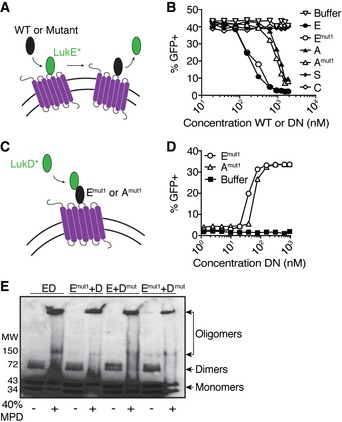
Schematic depicting competition assay using GFP‐LukE and unlabeled WT or mutant toxins.
Competition assay using GFP‐LukE (312.5 nM) and a titration of unlabeled WT or dominant‐negative (DN) toxin subunits evaluated by flow cytometry on the surface of primary human PMNs. E = LukE, Emut1 = LukEmut1, A = HlgA, Amut1 = HlgAmut1, S = LukS‐PV, C = HlgC. Data are presented as total fluorescence emitted from PMNs. A representative of three independent experiments each done with PMNs isolated from different donors is shown.
Schematic depicting recruitment assay using GFP‐LukD and unlabeled WT or dominant‐negative toxins.
Recruitment assay using GFP‐LukD (156.3 nM) and a titration of unlabeled dominant‐negative (DN) toxin subunits evaluated by flow cytometry on the surface of primary human PMNs. Emut1 = LukEmut1, Amut1 = HlgAmut1. Data are presented as total fluorescence emitted from GFP‐positive PMNs. A representative of three independent experiments each done with PMNs isolated from different donors is shown.
Immunoblot using anti‐His antisera to detect dimer and oligomer formation by purified polyhistidine (6xHis)‐tagged WT or dominant‐negative LukED toxins in the absence or presence of 2‐methyl‐2,4‐pentanediol (MPD, 40% v/v). Emut1 = LukEmut1, Dmut = LukDmut. A representative of two independent experiments is shown.

