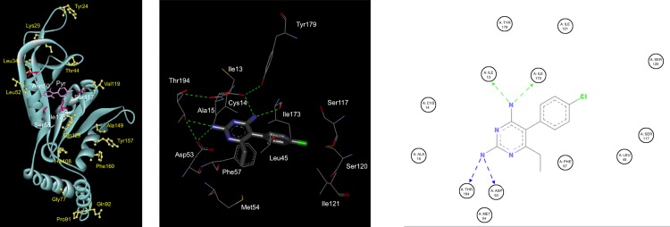
Fig 3. 3D structural model of the pkdhfr 91P mutation and molecular docking of pyrimethamine in the active site.
*Image panel attached separately. a) 3D structural model of pkdhfr with pyrimethamine bound at the active site. Mutations found in this study were shown as ball and stick coloured yellow. Equivalent residues of pkdhfr known to be related to pyrimethamine resistance in pfdhfr were presented as stick coloured dark pink and pyrimethamine shown as stick coloured magenta. b) Modelled pkdhfr binding site interaction with pyrimethamine. Pyrimethamine molecule was presented as stick and amino acid residues of pkdhfr were presented as line with carbon, nitrogen, oxygen and chloride colored as dark grey, blue, red and green, respectively. Hydrogen bonds are shown as green dashed line. c) 2D ligand interaction diagram of modelled pyrimethamine binding with all surrounding residues in active site of pkdhfr showing only contact with Ile13, Asp53, Ile 173 and Thr194. Hydrogen bonds are shown as dashed line. Figure was generated by Discovery Studio Visualizer–Accelrys.

