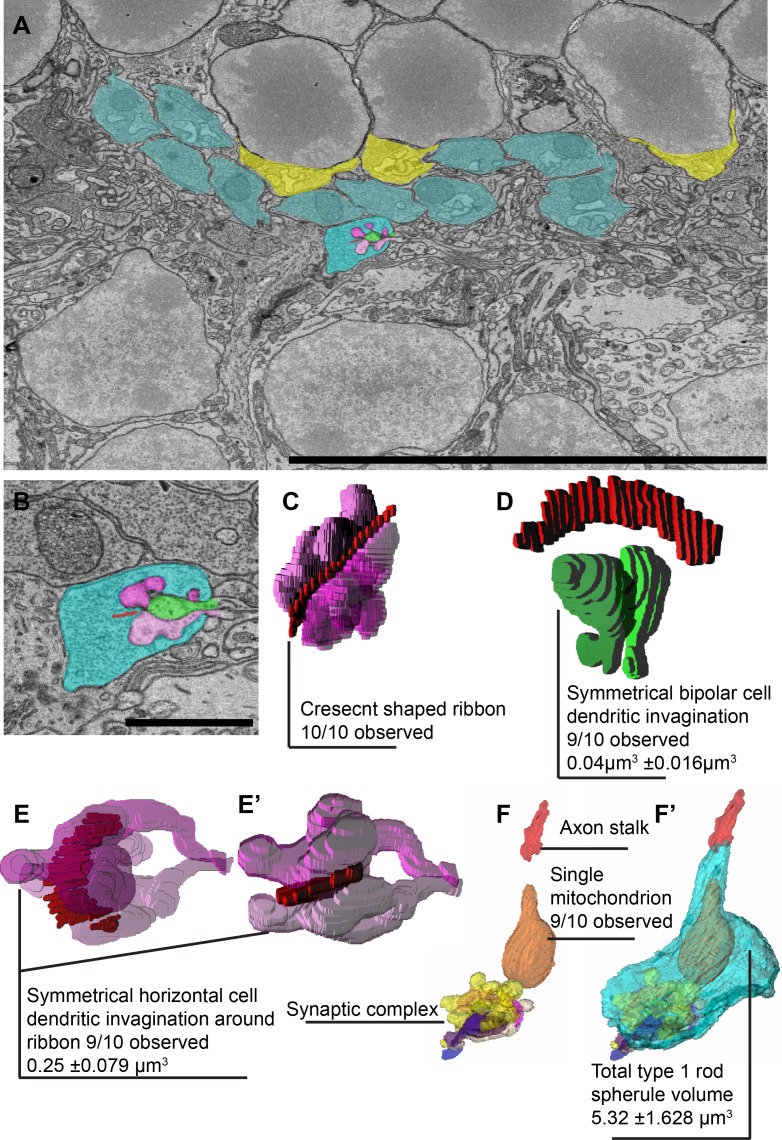
Fig 2. Internal structure of a stereotypical R1 rod spherule.
A, EM image of WT retina focused on the outer plexiform layer (OPL). R1 rod spherules are colored in blue and R2 rod spherules are colored in yellow. The center spherule is highlighted for reconstruction. B, High magnification of the EM image with each intracellular structure colored (red is the ribbon, green is invaginating bipolar cell dendrites, purple is invaginating horizontal cell axons). C, 3D reconstruction of R1 ribbon and invaginating horizontal cell axons. D, 3D reconstruction of R1 ribbon and invaginating bipolar cell dendrites. Note the symmetry of the bipolar dendrites. E and E’, Ribbon with horizontal cell axons from a different angle show the two invaginating axon tips maintain a spatial symmetrical orientation. F and F’, R1 rod spherule reconstruction modeled without and with cytoplasm region to show internal structures. (Scale bar in A is 10 μm, in B is 1.6 μm).

