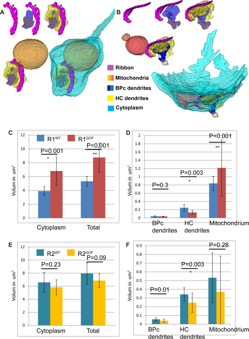
Fig 6. R1 spherules in DscamGoF retina are significantly different from R1 spherules in WT, while R2 spherules in DscamGoF do not differ significantly from their WT counterparts.
A and B, Reconstruction of R1 and R2 spherule in DscamGoF retina. C and D, Statistical analysis of R1 spherules between WT and DscamGoF retina show significant differences in all but invaginating bipolar cell dendrite volume. E and F, Statistical analysis of R2 spherules comparing WT and DscamGoF retina show insignificant differences in all but invaginating bipolar cell dendrite volume.

