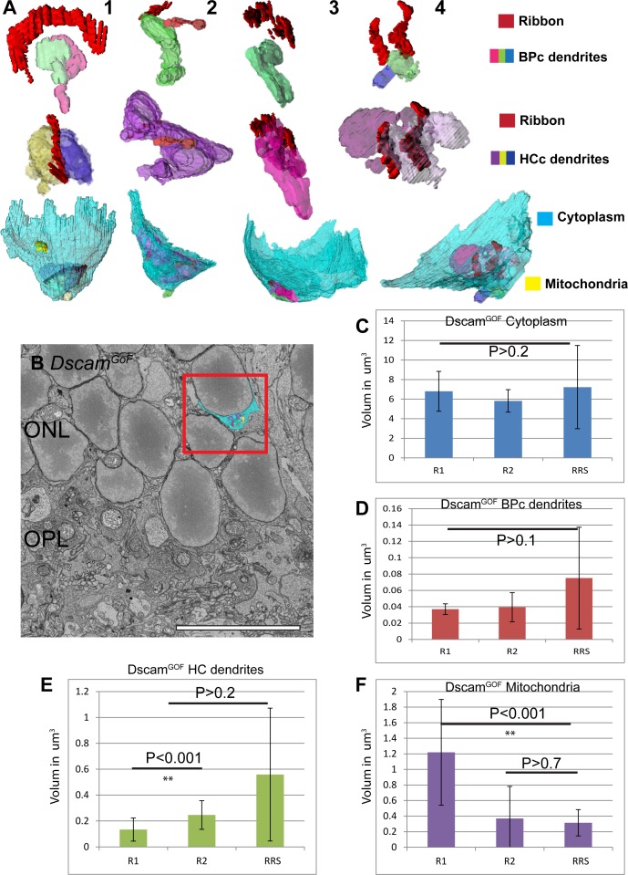
Fig 7. Retracted spherules in DscamGoF are similar to R2 spherules but with altered internal structures.
A, Reconstruction of retracted rod spherules (RRS) in DscamGoF retina. Only one spherule shows normal morphology and the rest display different degrees of abnormal morphology in the size and orientation of the synaptic complex. B, Retracted spherules in DscamGoF retina were identified greater than one rod soma deep into the ONL. C, Statistical analysis of spherules types in DscamGoF retina revealed no difference in cytoplasm volume across spherule types. D, Statistical analysis of spherules types in DscamGoF retina revealed no difference in invaginating bipolar cell dendrite volume across rod types. E, Statistical analysis of spherule types in DscamGoF retina show significant differences between horizontal cell axon volume in R1 and R2 spherules but not compared to retracted spherules, likely due to the high variance in horizontal cell axon volume. F, Statistical analysis of mitochondrial volume across spherule types in DscamGoF retina show significant differences between R1 and other spherules (Bar in B is 10μm).

