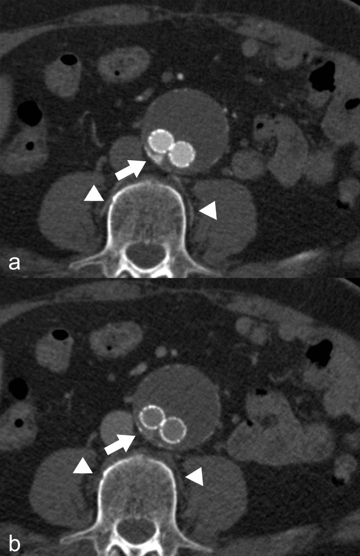
Fig 1. Feeding vessel detection in arterial and venous phase acquisition.
(a) Arterial phase acquisition: Clearly perceptible blush of contrast agent in the aneurysm sac (arrow) indicative of the presence of a type 2 endoleak. Consecutive, feeding lumbar arteries of this segment show the same contrast as the aorta (arrowheads). (b) Venous phase acquisition: Similarly, clearly detectable type 2 endoleak (arrow). However, potentially feeding lumbar arteries show no contrast enhancement (arrowheads).

