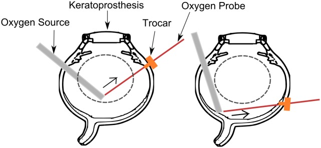
Fig 2. Ex vivo porcine eye preparation and intravitreal oxygen measurement methods.
Left: Oxygen source is positioned in the mid-vitreous and the oxygen probe is retracted in the direction shown by the arrow. Right: Oxygen source is positioned in contact with the retinal tissue and the oxygen probe is retracted in the direction shown by the arrow. The probe is positioned such that it stays in the posterior vitreous region. The trocar is used to facilitate oxygen probe entry and retraction without creating any motion artefact. The dashed lines indicate regions that are designated has mid vitreous and posterior vitreous.

