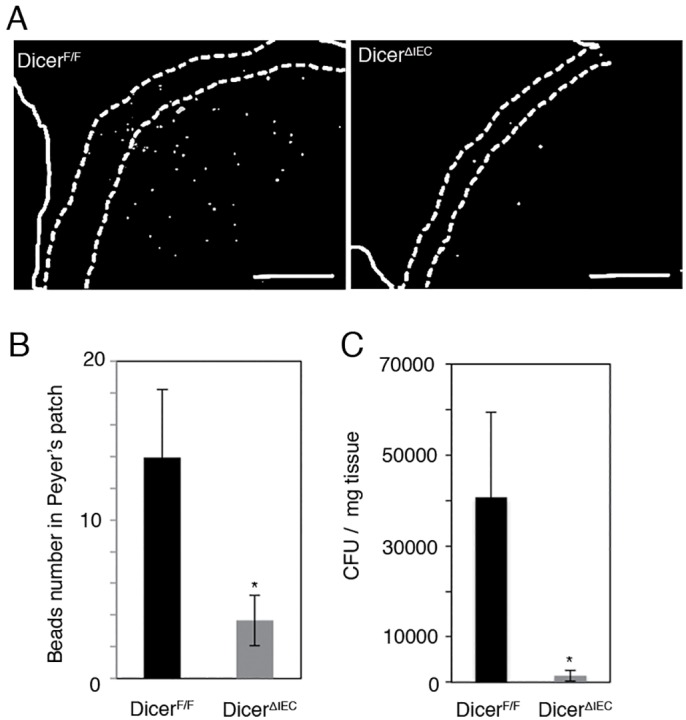
Fig 5. Impaired antigen uptake by DicerΔIEC M cells.

(A) DicerΔIEC and DicerF/F mice were inoculated by gavage with 1 x 10¹¹ FluoSpheres. After 4 hours, frozen sections were prepared to examine translocated beads in PPs. Scale bars: 100 μm (B) Count data of beads taken up in PP each mouse strain. Data are expressed as the mean ± SE of four different samples for each group. **P < 0.01. (C) DicerΔIEC and DicerF/F mice were inoculated intragastrically by gavage with 1 x 10⁸ CFU of Yersinia enterocolitica. After 24 hours, the bacterial translocation to Peyer’s patches was examined by plating PP homogenates. Data are expressed as the mean ± SE of five different mice/each group. *P < 0.05.
