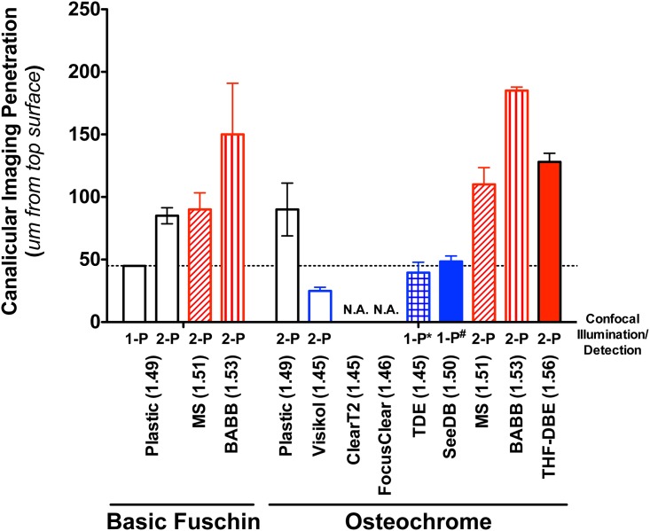
Fig 6. Quantification of bone’s canalicular confocal imaging penetration following en bloc staining and optical clearing.
Non-aqueous clearing agents (MS, BABB, and THF-DBE) showed an appreciable ability to improve the visualization of fine canalicular detail (features on the order of 500 to 1000-nm in diameter) at depths 2 to 4-times that of the traditional “gold-standard” technique (plastic embedded 1-photon). Data represent the mean ± SD depth at which imaging of discrete canalicular structure is lost in stained, cleared, and imaged samples (n = 3–4 samples per clearing agent). 1-P indicates 1-photon imaging. 2-P indicates 2-photon imaging. ClearT2 and FocusClear were incompatible with Osteochrome staining, while TDE and SeeDB were incompatible with 2-photon imaging (*). Statistical analyses were not performed given the subjective nature of defining canalicular imaging penetration depth.

