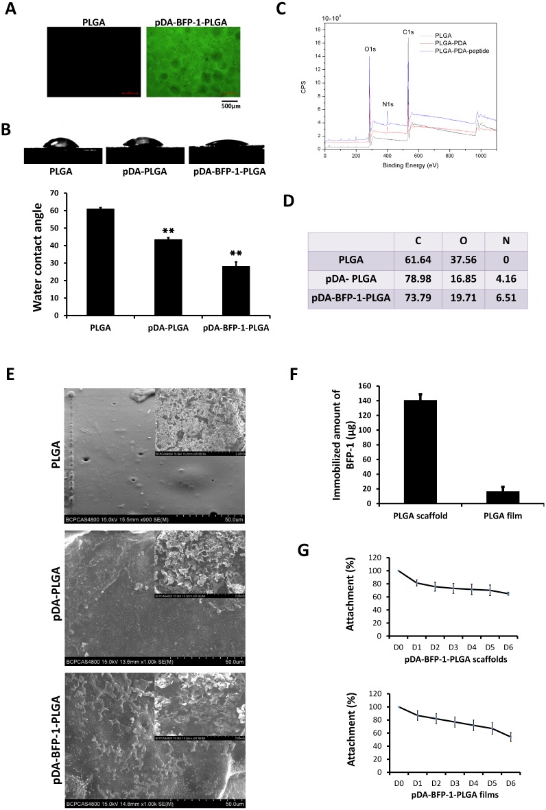
Fig 3. Surface characterization of engineered PLGA substrates.
(A) Fluorescent images of PLGA substrates with (right) or without (left) immobilized, fluorescently labeled peptides (FITC-BFP-1). (B) Contact angle measurement of PLGA, pDA-PLGA, and pDA-BFP-1-PLGA films (**: p <0.01). (C) X-ray photoelectron spectroscopy (XPS) analysis of PLGA, pDA-PLGA and BFP-1-pDA-PLGA films. (D) Chemical composition of film surfaces. (E) Scanning electron microscopy of PLGA, pDA-PLGA and pDA-BFP-1-PLGA scaffolds. (F) Amount of immobilized BFP-1 (μg). (G) Release curves of pDA-BFP-1-PLGA scaffolds and films.

