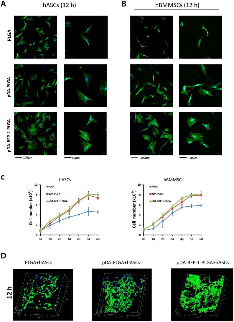
Fig 4. Adhesion and proliferation of hASCs and hBMMSCs on PLGA films.
hASCs: human adipose-derived stem cells; hBMMSCs: human bone marrow-derived mesenchymal stem cells. Confocal micrographs of hASCs (A) and hBMMSCs (B) on PLGA, pDA-PLGA and pDA-BFP-1-PLGA films after culturing for 12 h. Phalloidin is colored green and nuclei are colored blue. (C) Proliferation curves of hASCs and hBMMSCs on PLGA, pDA-PLGA and pDA-BFP-1-PLGA film surfaces. (D) Confocal micrographs (50x) of hASCs on PLGA, pDA-PLGA and pDA-BFP-1-PLGA scaffolds after culturing for 12 h. Phalloidin is colored green and nuclei are colored blue. The green signal shown in these pictures indicate the volume of cells.

