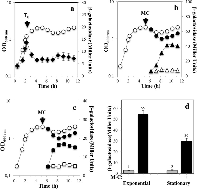
Fig 2. Levels of ß-galactosidase from recA-lacZ in growth and sporulation and with and without DNA damage.
B. subtilis strain YB3001 containing a recA-lacZ fusion was grown and induced to sporulate in DSM (a-c) The optical densities of cultures were measured without (○) or after (●) DNA damaging treatment. Samples were also collected at different times during growth and sporulation and were processed and assayed for ß-galactosidase specific activity. In (a) ß-galactosidase from recA-lacZ was assayed throughout growth and sporulation (◆). In (b,c) 4 h after the onset of sporulation (T0), the culture was divided into two subcultures; one subculture was challenged with M-C (500 ng/mL) and the other one was untreated. Cells samples from untreated (open symbols) or treated (filled symbols) were collected at the indicated times and ß-galactosidase specific activity in the mother cell (b, triangles) and forespore (c, squares) fractions was determined, all as described in Materials and Methods. In (d), B. subtilis YB3001 was propagated in PAB medium, when the culture reached an OD600nm = 0.5 (Exponential) or 4 h after T0 (Stationary), vegetative cells were treated (black bars) or not (gray bars) with M-C (500 ng/mL) for 1.5 h and then the cultures were processed for determination of ß-galactosidase as described above. Results are the average of values from three independent experiments ± standard deviations (SD) of ß-galactosidase specific activity.

