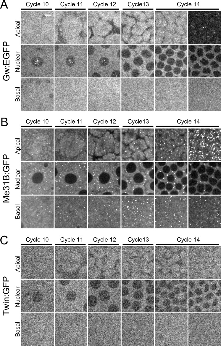
Fig 1. Cytoplasmic GW-bodies are absent during early embryonic development and form de novo at cellularization.
Live imaging of Gw:EGFP, Me31B:GFP, and Twin:GFP during cortical syncytial cycles and cellularization. Apical, nuclear, and basal images of embryos during cycles 10, 11, 12, 13, and 14. Cycle 14 images were taken just after completion of cycle 13 (left image) or during cellularization (right image). (A) Embryos expressing Gw:EGFP. Gw is present in the nucleus during cortical syncytial cycles. Cytoplasmic GW-bodies form primarily during cellularization. (B) Embryos expressing Me31B:GFP. Me31B labeled P-bodies are present throughout cortical syncytial cycles. New P-body structures form in large numbers during cellularization. (C) Embryos expressing Twin:GFP. Twin is not associated as punctate structures. Scale bar = 5 μm.

