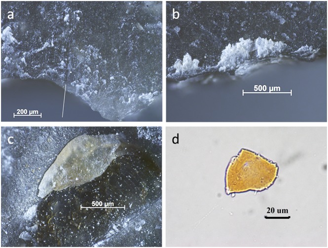
Fig 3. Residues identified on BT6 (unused) as described during blind test.
a) White use-residue with evident directionality (indicated by arrow) along Edge A of the ventral surface; b) high magnification image of white, amorphous residue on tool edge; c) possible fatty deposits (beneath scale bar); d) collagen residue stained with Orange G, as viewed under transmitted light.

