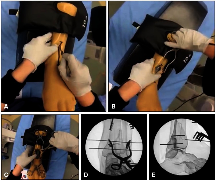
Fig. 1A–E.
(A–C) The articular fracture reduction model is shown at various stages in the exercise, revealing the radioopaque surrogate bone specimen with soft tissue sleeve. (D) AP and (E) lateral fluoroscopic images of the intraarticular distal tibia fracture are shown, nearing final reduction with Kirschner wires having been placed by the resident.

