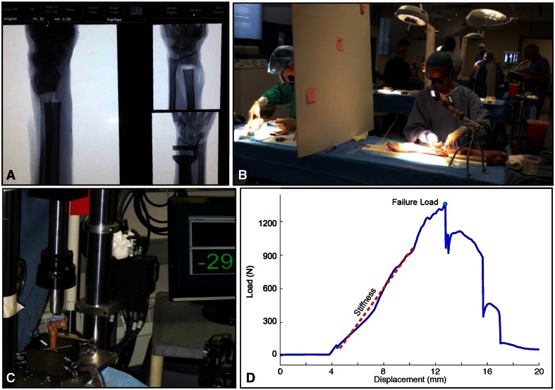
Fig. 3A–D.
This montage depicts the articular fracture simulation involving the fixing of a simulated extraarticular fracture of the distal radius in an upper extremity cadaver specimen. (A) The radiographic images in the upper left show the osteotomy with the cutting jig immediately proximal to the distal radioulnar joint (DRUJ) shown in the lower right. (B) Three residents are taking the examination at the same time here. A faculty member is grading one resident in the background. The faculty grading the residents in the foreground have stepped out of the picture, and a C-arm available to the residents is seen in the background. (C) This image shows the loading of the fracture fixation construct. (D) This illustrative displacement versus force data tracing shows load uptake and the basis for assessing stiffness and failure load.

