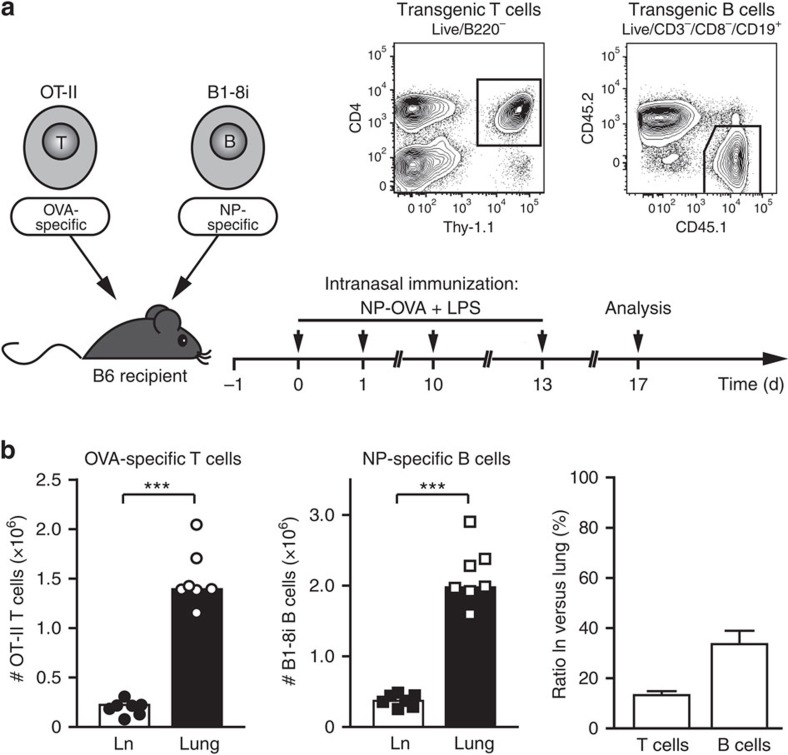
Figure 1. Inflamed lung tissue is the major reservoir of antigen-specific T and B cells.
(a) General set-up for the airway inflammation model. Preactivated OT-II T cells and naive B1-8i/kappa light chain knockout B cells were transferred into syngeneic C57BL/6 mice. OT-II cells express a T-cell receptor specific for OVA, B1-8i cells have a knock-in for the germline VH186 heavy chain, which recognizes the hapten NP in combination with any lambda 1 light chain31. On the indicated days, recipient mice were immunized i.n. with an NP–OVA conjugate and LPS. Cells from lungs and lung-draining lymph nodes were typically analysed on day 17 when airway inflammation was fully established. Antigen-specific T and B cells were identified by flow cytometry using the congenic markers Thy-1.1 and CD45.1, respectively. (b) Total numbers of antigen-specific T and B cells in lung-draining lymph node (Ln) and lung tissue on day 17. Shown are absolute cell numbers of a representative experiment and the ratio of antigen-specific T and B cells in lymph node versus lung (pooled data from 6 experiments with 34 animals). Error bars show s.e.m. ***P<0.001 by Mann–Witney U-test.

