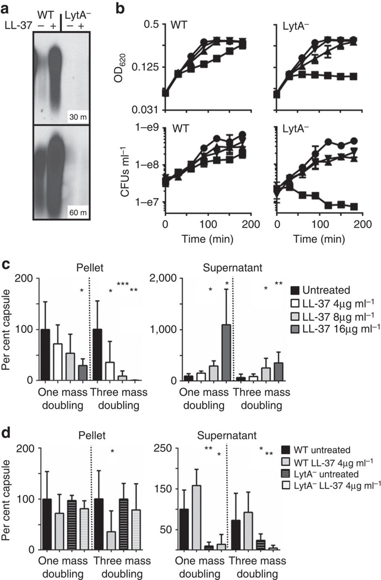
Figure 2. Role of LytA autolysin in capsule shedding.
(a) WT TIGR4 or the isogenic deletion mutant in the lytA gene (LytA−) were treated with 4 μg ml−1 LL-37 in a capsule-shedding assay for 30 or 60 min as indicated. The supernatants were then analysed by capsule blot. (b) TIGR4 (WT) or the mutant in lytA (LytA−) were treated at t=0 with LL-37 at 16 μg ml−1 (squares), 8 μg ml−1 (triangles), 4 μg ml−1 (inverted triangles) or untreated (circles). Cultures were then monitored by optical density and samples collected for enumeration of viable CFUs at the indicated times. (c) TIGR4 (WT) was grown in C+Y to OD 0.4. Cultures were then back diluted to OD 0.2 (1 mass doubling) or OD 0.05 (three mass doublings) and LL-37 was added at the indicated concentrations. At OD 0.4 the cultures were harvested and pellet and supernatant fractions were analysed by capsule blot. Relative capsule amounts were calculated by comparison with a purified capsule standard and were normalized to untreated cells for each fraction. The results are the mean and s.d. from three independent experiments (100% pellet: 55.9 μg ml−1±32.1 n=3; supernatant 2.68 μg ml−1±0.827, n=3). (d) The LytA− strain was grown as in c and treated with 4 μg ml−1 LL-37 where indicated. For reference, the results for WT in the same conditions in c were compared and plotted against the LytA− results. *P<0.05 **P<0.01 ***P=0.001 unpaired t-test with Welch's correction.

