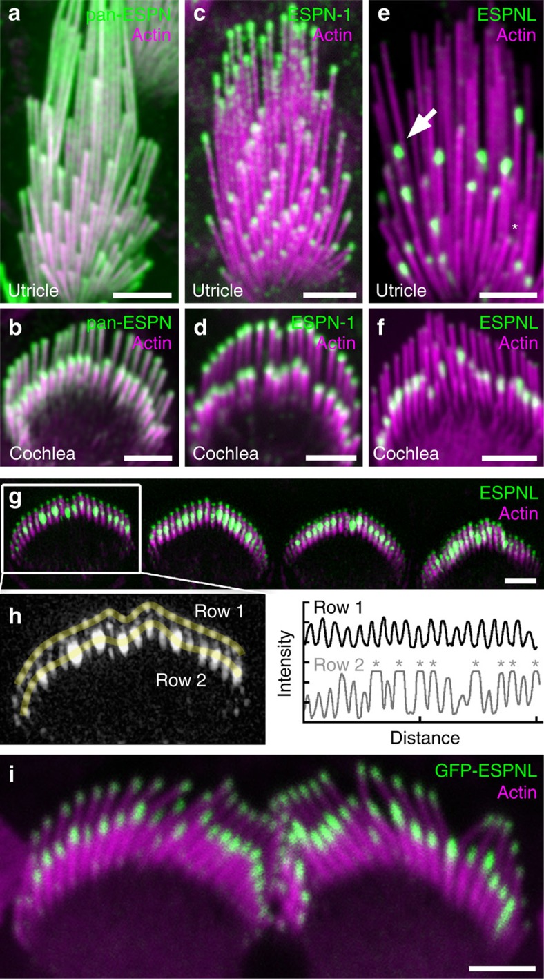
Figure 3. Immunolocalization of ESPN-1 and ESPNL.
(a,b) Pan-ESPN antibody (green) and actin (magenta) labels utricle (a) and cochlear outer hair cell (b) stereocilia throughout. (c,d) ESPN-1 antibody labels the tips of utricular (c) and cochlear (d) stereocilia. (e) ESPNL antibody (ab170747) labels some stereocilia tips strongly (arrow), some weakly (asterisk), but does not label tallest stereocilia of utricle hair cell. (f) ESPNL antibody labels most but not all stereocilia tips of row 2 in bundle of outer hair cell. (g) Structured illumination microscopic image of ESPNL labelling (BG35961) of inner hair-cell bundles. (h) Quantification of ESPNL labelling of one hair bundle from (G). (left) Yellow lines indicate quantification trajectories; and (right) uniform labelling in row 1 but saturation of some tip signals in row 2 (asterisks). (i) GFP-ESPNL labelling of P0.5 inner hair cells following in utero electroporation at E12. Scale bars, 2 μm.

