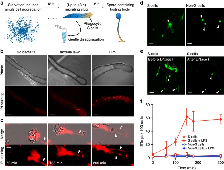
Figure 1. Bacteria or LPS stimulate S cells to produce ETs.
(a) Development of D. discoideum from single cells to a multicellular organism. The black dots in the migrating slug indicate S cells. (b) Red fluorescent dots (indicative of extracellular DNA) from AX4 slugs migrating on PI-containing agar plates in absence or presence of K.p. or K.p. LPS. (c) Disaggregated AX4 slug cells release ETs (arrowheads, PI staining) on LPS stimulation. See also Supplementary Movie 1. (d) On LPS stimulation, DNA fibres (arrowheads, SYTO9 staining) were released from FACS-purified S cells, but not from non-S cells. (e) ETs disappeared after 10-min exposure to DNase I, while the protected nuclear DNA remains visible (arrowheads). (f) Quantification of ETs production from purified S and non-S cells with or without LPS exposure, n=3. Error bars, s.e.m. Scale bars, (b) 100 μm; (c–e) 10 μm.

