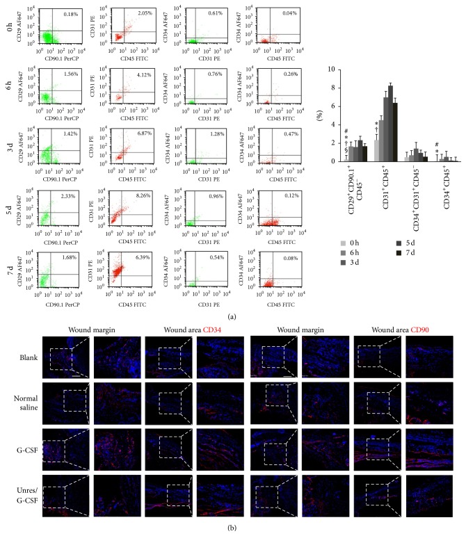
Figure 1.
Elevated MSCs and EPCs anticipated wound healing in the hemorrhagic shock rats. (a) Representative density plots of the alterations of the BMSC distribution in the circulating blood at normal state (0 h), 6 h, 3 d, 5 d, and 7 d after G-CSF mobilization and HHES resuscitation. The typical cell surface markers represented as MSCs (CD45−CD29+CD90+), HSCs (CD45+CD31+ or CD45+CD34+), and EPCs (CD45−CD31+CD34+). # P < 0.05 versus the 6 h values; ∗ P < 0.05 versus 3 d values; † P < 0.05 versus 5 d values; § P < 0.05 versus 7 d values. (b) Representative images showed that CD34 (left, red) and CD90 (right, red) positive cells located both at the wound margins and wound areas at 6 h. Scale bar = 200 μm. Each experiment was repeated three times and typical pictures were shown.

