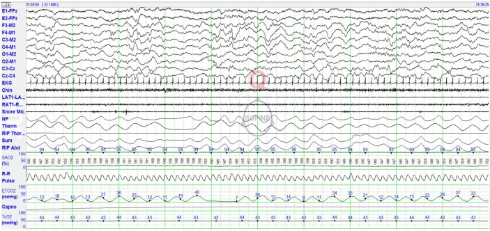
Figure 2. Polysomnographic (PSG) montage for recording infants 0–2 mo of age.
Eye channels (left eye [E1], right eye [E2]) are referenced to the midline frontopolar electrode (FPz). Electroencephalogram (EEG montage is left frontal-right mastoid (F3-M2), right frontal-left mastoid (F4-M1), left central-right mastoid (C3-M2), right central-left mastoid (C4-M1), left occipital-right mastoid (O1-M2), right occipital-left mastoid (O2-M1), left central-midline central (C3-CZ), and midline central-right central (CZ-C4). This EEG montage allows easy recognition of low voltage 12–14 Hz sleep spindles, which may be seen as early as 43 w, usually present by 45–48 w CA. Other PSG signals recorded include: EKG, chin electromyogram (EMG), left and right anterior tibialis (L Leg, R Leg) muscles, snore microphone, nasal pressure (NP), thermal sensor, respiratory inductance plethysmography (RIP) thoracic, RIP abdomen, RIP SUM, pulse oxygen saturation (SpO2), R-R interval, pulse waveform, endtidal carbon dioxide (EtCO2), capnogram, and transcutaneous carbon dioxide (tcCO2).

