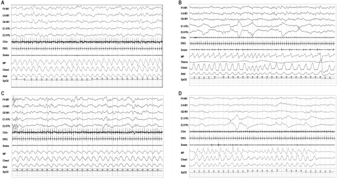
Figure 4. Representative examples of polysomnographic patterns of sleep in infants 0–2 mo of age.
(A) 30-sec epoch of rapid eye movement (REM) sleep with mixed (M) electroencephalogram (EEG) pattern, REMs and low chin electromyogram (EMG) tone typically seen following a period of wakefulness; (B) 30-sec epoch of NREM sleep with high voltage slow (HVS) EEG pattern, no eye movements, preserved chin EMG, and regular respiration, which often precedes longer period of trace alternant (TA) in infants at 38–42 w conceptual age (CA); (C) 30-sec epoch of nonrapid eye movement (NREM) sleep with trace alternant (TA) EEG pattern, no eye movements, regular respiration; and (D) 30-sec epoch of REM sleep with a low voltage irregular (LVI) EEG pattern, rapid eye movements (REMs), irregular respiration, and low chin EMG tone.

