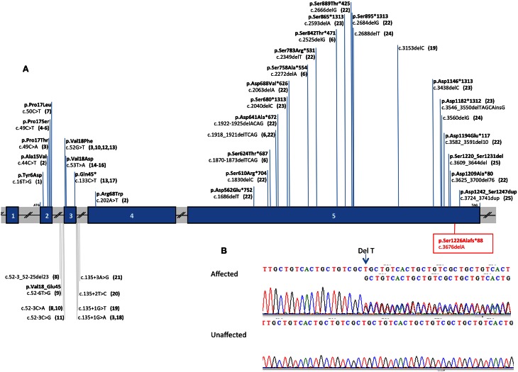
Figure 3.
DSPP mutation analysis. DSPP mutations are indicated against the gene structure (reference sequence: NM_014208.3). (A) Schematic representation of DSPP: this gene contains 5 exons (vertical blue hatches), the position of the start codon (ATG) and the stop codon (TAG) are indicated respectively in exon 2 and exon5. The known mutations in the DSPP gene are summarized against the gene structure and associated to a literature reference. The new mutation described in this paper is boxed and written in red. For example: the most 5′ DSPP mutation, near the initiation codon (ATG) is lying in exon 2 and described as a single nucleotide variant c.16T>G leading to the following amino acid changes in the protein p.Tyr6Asp and reported in the literature in quoted reference (Rajpar et al., 2002). (B) Electrophoregrams of a part of DSPP exon 5 showing the heterozygous mutation in an affected person and the normal sequence in an unaffected individual. The deletion of 1T is indicated with an arrow, this deletion creates a shift in the reading frame in position 3676 of the cDNA reference sequence, resulting in 2 superposed sequences. On the scheme the numbering corresponds to the following references: 1. Rajpar et al. (2002); 2. Malmgren et al. (2004); 3. Xiao et al. (2001); 4. Zhang et al. (2007) and Qu et al. (2009); 5. Hart and Hart (2007); 6. Mcknight et al. (2008a); 7. Li et al. (2012) and Lee et al. (2013); 8. Wang et al. (2009); 9. Lee et al. (2008); 10. Holappa et al. (2006); 11. Kim et al. (2004); 12. Kim et al. (2005); 13. Song et al. (2006); 14. Lee et al. (2011b); 15. Kida et al. (2009); 16. Lee et al. (2009); 17. Zhang et al. (2001); 18. Wang et al. (2011); 19. Mcknight et al. (2008b); 20. Zhang et al. (2011); 21. Bai et al. (2010); 22. Nieminen et al. (2011); 23. Song et al. (2008); 24. Lee et al. (2011a); 25. Dong et al. (2005).

