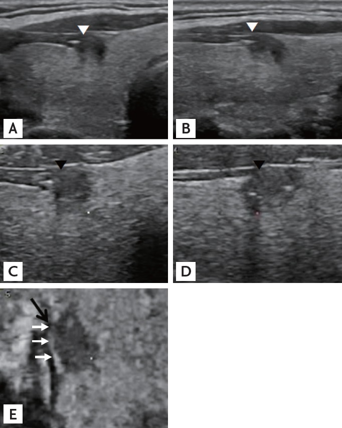
Figure 4.

A representative presence of both contact and protrusion (C1P1) papillary thyroid carcinoma. The carcinoma is in contact with the thyroid capsule and protrudes into adjacent structures in the transverse (A) and longitudinal (B) planes on sonograms from two-dimensional ultrasonography (white arrowheads). Tomographic ultrasound imaging rendered images from three-dimensional ultrasonography also demonstrate that the carcinoma is in contact with and protrudes in the transverse (C) and longitudinal (D) planes (black arrowheads). In the coronal (E) plane, the lesion is in contact with the capsule (white arrows), but protrusion is not definite (black arrow). Final pathologic finding was negative for extrathyroidal extension.
