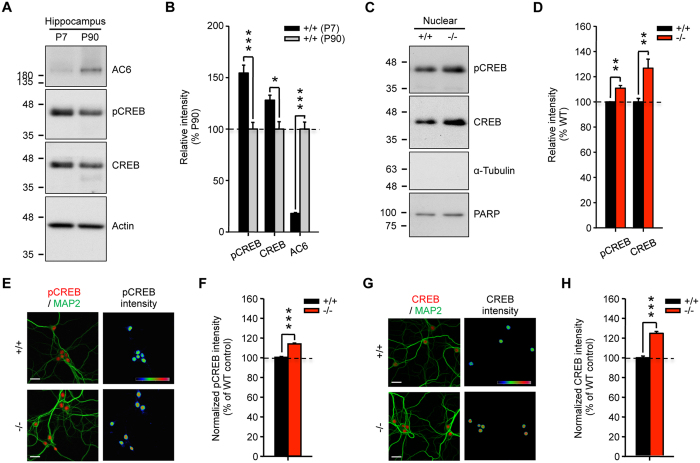
Figure 1. AC6 negatively regulates CREB level in the hippocampus.
(A,B) Total lysate (Total, 20 μg) harvested from the hippocampi of postnatal day (P) P7 and P90 (n = 3–6) mice were subjected to WB analysis. (A) The expression level of AC6, pCREB, CREB, and actin were detected using the indicated antibody. Actin was used as a loading control. The level of the indicated protein was quantified, normalized with that of P90, and shown in (B). (C,D) Hippocampal nuclear fraction (5 μg) of AC6+/+ (n = 6) and AC6−/− mice (n = 6) at P90 (3 months of age) were subjected to WB analysis. (C) The expression level of pCREB and CREB were detected using the indicated antibodies. PARP was used as the internal loading control of nuclear fraction. The protein expression level was quantified and is shown in (D). (E–H) Primary hippocampal neurons of AC6+/+ and AC6−/− mice were fixed at DIV14 to analyze the levels of pCREB (red, E) and CREB (red, G) by immunostaining. (E,G) MAP2 (green) is a neuronal marker. The color bars in the upper-right panel, from cold to warm colors, represent low to high fluorescence intensities of pCREB or CREB as indicated. Scale bars, 25 μm. The intensity of pCREB (F; AC6+/+, n = 436; AC6−/−, n = 487) or CREB (H; AC6+/+, n = 157; AC6−/−, n = 139) in each cell was divided by the mean value of the AC6+/+ cells and is represented as the means ± SEM. Data were analyzed using Student’s t-test. *p < 0.05; **p < 0.01; ***p < 0.001, versus the control group.

