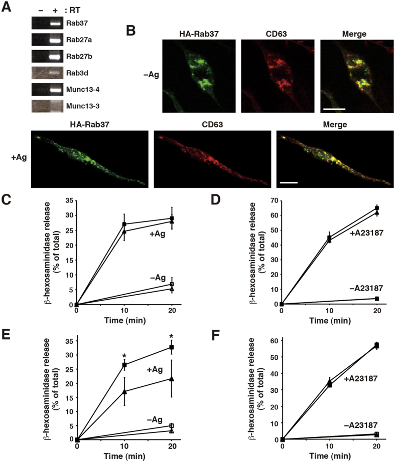
Figure 1. The Rab37 GTPase resides on secretory granules and its dominant-active mutant shows increased antigen-induced degranulation in RBL-2H3 cells.
(A) Endogenous Rab37 expression. Total RNA from the cells was subjected to first-strand cDNA synthesis with (+) or without (−) reverse transcriptase (RT), followed by PCR amplification with specific primer sets for the indicated genes. PCR products were separated on agarose gel and stained with ethidium bromide. The full-length gel images are presented in Supplementary Fig. S1. (B) Intracellular localisation of Rab37. Cells transfected with the HA-Rab37 plasmid were sensitised with IgE and stimulated with (+Ag) or without (−Ag) antigen in a Ca2+ -free medium, followed by fixation and co-immunostaining with anti-HA and anti-CD63 antibodies. Merged images are also shown on the right. Note that panels (−Ag) are enlarged images of the cell focused on perinuclear region with parts of unstained rod-like protrusions and that panels (+Ag) are images of the whole cell. Bars, 15 μm. (C) Antigen-induced degranulation in the cells expressing exogenous Rab37. G418-surviving cells transfected with the HA-Rab37 (square, n = 4) or the control (triangle, n = 4) plasmid were sensitised with IgE and stimulated with (+Ag) or without (−Ag) antigen for the indicated time periods. (D) A23187/TPA-induced degranulation in the cells expressing exogenous Rab37. The G418-surviving transfectants described in (C) were stimulated with (+A23187) or without (−A23187) A23187 together with 12-O-tetradecanoylphorbol-13-acetate (TPA) for the indicated time periods. (E) Antigen-induced degranulation in the cells expressing a dominant-active Rab37 mutant. G418-surviving cells transfected with the HA-Rab37DA (square, n = 4) or the control (triangle, n = 4) plasmid were sensitised and stimulated as in (C). (F) A23187/TPA-induced degranulation in the cells expressing the dominant-active Rab37 mutant. The G418-surviving transfectants described in (E) were stimulated as in (D). In (C–F) β-hexosaminidase activity was measured in the supernatant and the cell lysate, and the degree of release was expressed as a percentage of the total activity. *p < 0.05, compared to the stimulated control transfectant.

