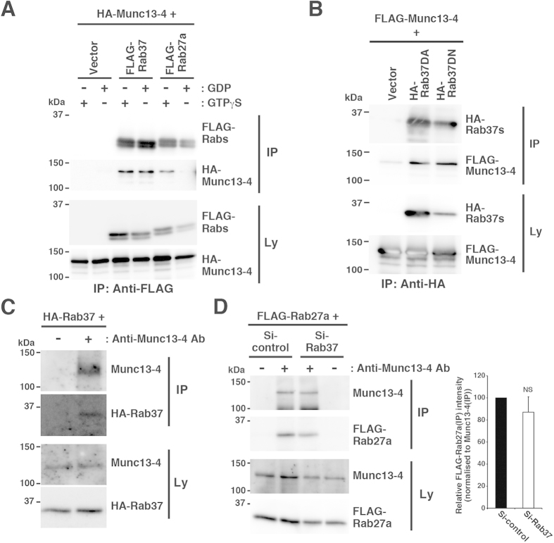
Figure 5. Rab37 interacts with Munc13-4.
(A) Rab37-Munc13-4 interaction in COS7 cells. Lysates were prepared from the cells transfected with HA-Munc13-4 plasmid together with control (Vector), FLAG-Rab37, or FLAG-Rab27a plasmid in the presence (+) or absence (−) of GTPγS or GDP and subjected to immunoprecipitation with anti-FLAG antibody. The lysates (Ly) and immunoprecipitates (IP) were then subjected to western blot analyses with anti-HA (3F10) or anti-FLAG antibody. (B) Munc13-4 interacts with both the dominant active and the dominant negative mutants of Rab37. Lysates were prepared from COS7 cells transfected with FLAG-Munc13-4 plasmid together with control (Vector), HA-Rab37DA, or HA-Rab37DN plasmid and subjected to immunoprecipitation with anti-HA antibody. The lysates (Ly) and immunoprecipitates (IP) were analysed as in (A). (C) Rab37-Munc13-4 interaction in RBL-2H3 cells. Cells were transfected with the HA-Rab37 plasmid and cultured in the presence of G418 for 24 h. The surviving cells were treated with dithiobis succinimidyl propionate (DSP), lysed, and subjected to immunoprecipitation with (+) or without (−) anti-Munc13-4 antibody (Anti-Munc13-4 Ab). The lysates (Ly) and immunoprecipitates (IP) were then subjected to western blot analyses with anti-HA (3F10) or anti-Munc13-4 antibody. (D) Rab27-Munc13-4 interaction in the Rab37-knockdown RBL-2H3 cells. Cells were transfected with FLAG-Rab27a plasmid, together with Si-control or Si-Rab37 plasmid and cultured in the presence of G418 for 24 h. The surviving cells were treated with DSP, lysed in the presence of GTPγS, and subjected to immunoprecipitation with (+) or without (−) anti-Munc13-4 antibody (Anti-Munc13-4 Ab). The lysates (Ly) and immunoprecipitates (IP) were then subjected to western blot analyses with anti-FLAG or anti-Munc13-4 antibody. The graph represents the relative quantity of FLAG-Rab27a co-immunoprecipitated with Munc13-4, with the quantity in the Si-control transfectant set to 100. Bars represent mean ± SD (n = 4). NS, not significant. The full-length blotting images of (A–D) are presented in Supplementary Figs S13, S14, S16 and S17, respectively.

