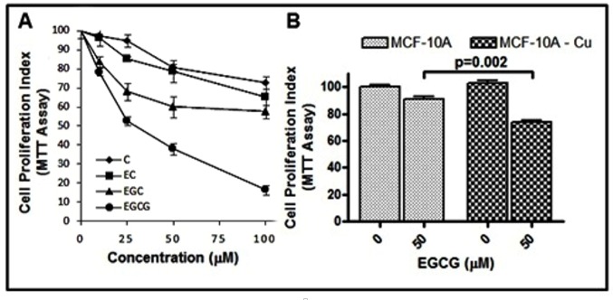
Figure 11.
(A) The effects of C, EC, EGC and EGCG on the growth of MDA-MB-231 breast cancer cells as detected by MTT assay. The cells were incubated with indicated concentrations of catechins for 48 h, and the results are expressed relative to control (vehicle-treated) cells. (B) MCF10A (normal breast epithelial cells) and MCF10A+Cu (MCF-10A cells cultured in copper-enriched medium) were treated with either vehicle (0 μM) or 50 μM EGCG for 72 h.

