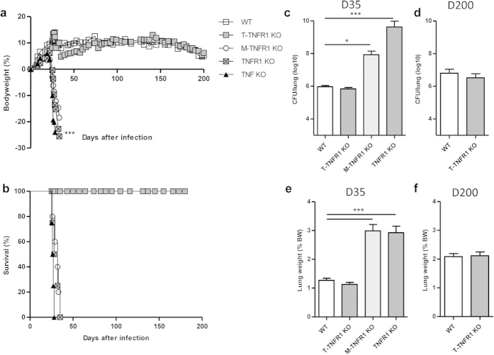
Figure 7. T-TNFR1 KO mice control acute M. tuberculosis infection.
Mice specifically deficient for TNFR1 in T cells (T-TNFR1 KO) or myeloid cells (M-TNFR1 KO), fully TNFR1 or TNFα deficient mice and wild-type mice were exposed to M. tuberculosis H37Rv (1000 CFU/mouse i.n.) and monitored for relative body weight gain (a) and survival (b). Pulmonary bacterial load (c,d) and lung wet weight (e,f) were measured at 35 days (c,e) and 201 days (d,f) post-infection. Results are expressed as mean +/− SEM from n = 9–12 mice per group pooled from 3 independent experiments up to day 35 and n = 9–10 from 2 experiments thereafter. *p < 0.05; **p < 0.01; ***p < 0.001.

