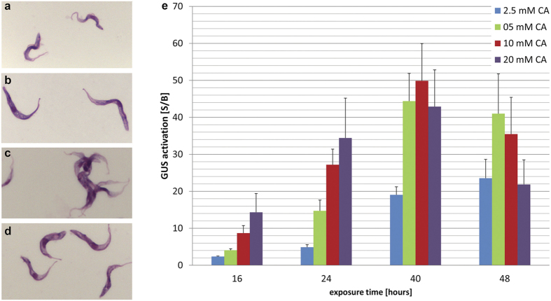
Figure 1. Establishment of GUS assay with CA.
Giemsa stained T. b. b. GUSone (a–d). Bloodstream forms (a), procyclic forms (b) parasites were differentiated to procyclics with a 24 h exposure to 5 mM CA at 27 °C followed by one week culture in SDM-79 medium at 27 °C. Bloodstream forms that were treated with 10 mM CA for 40 h at 37 °C (c), bloodstream forms that were treated with 10 mM CA for 40 h at 27 °C (d). GUS activation is dependent on CA concentration and exposure time at 37 °C (e). The starting density was 8 × 10⁵ parasites/ml for 16 h and 24 h, 5 × 10⁵ parasites/ml for 40 h, and 2.5 × 10⁵ parasites/ml for 48 h exposure. Bars indicate standard deviations of S/B from at least three independent assays. Cells exposed to CA at 37 °C tend to aggregate. CA = cis-aconitate.

